How XNAT is helping the NHS
The ICR/RM XNAT repository is a crucial component in the ongoing efforts to improve Radiology services within the Royal Marsden. We help clinical physicists test new imaging methodologies prior to introduction in the clinic. XNAT fulfils several important roles:
- organisation of imaging data into research projects;
- flexible display of data ordered by a variety of different criteria based on image metadata;
- provision of a web-accessible “single point of truth” for large multi-modal datasets;
- flexible zero-footprint image viewer;
- ability to add bespoke image manipulation tools not available via the RM Picture Archiving and Communication System (PACS);
- ability to annotate images and save the results in machine-readable formats for use in downstream processing.
CASE STUDY: AI-BASED RECONSTRUCTIONS IN MRI OF RECTAL CANCER
With increasing AI adoption in MR-reconstructions, robust quality assessment becomes paramount. While guidance exists saying institutions must verify and monitor AI-tools, there is scant literature offering practical measures for implementation. There is a need to develop efficient QC tests, which offer performance monitoring for AI-based reconstructions in diverse clinical settings.
This MRI Physics Team at RMH ran a study aimed at ensuring that AI-techniques meet clinical requirements at implementation and longitudinally.
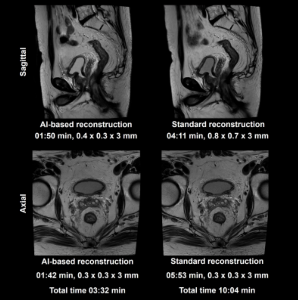
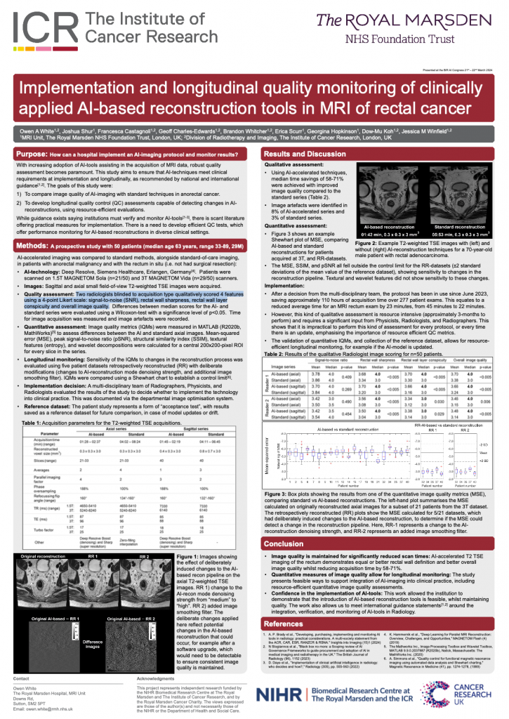
The project had two goals: (1) to compare image quality of AI-imaging with standard techniques in anorectal cancer; and (2) to develop longitudinal quality control (QC) assessments capable of detecting changes in AI-reconstructions without resource-intensive evaluations. A prospective study involving 40 patients utilised radiologist scoring and quantitative image-quality-metrics (IQMs). Retrospective reconstructions gauged sensitivity of IQMs to reconstruction pipeline changes.

Two radiologists, blinded to acquisition type. qualitatively scored four features (signal-to-noise (SNR), rectal wall sharpness, rectal wall layer conspicuity and overall image quality), using a four-point Likert scale. Results of the study were extremely encouraging. AI-reconstructions demonstrated 50-70% time savings with improved image quality. Feasibility of quantitative-IQMs for assessing AI-reconstructions was established, providing a practical solution for ongoing QC.
Key parts of this study would not have been possible without transferring data to the ICR/RM XNAT Image Repository and performing some of the initial evaluations (particularly the rectum protocol) using XNAT systems. In the words of the study lead author:
[This] just shows how useful XNAT is for clinical implementation, as well as research output.

The study had an immediate practical benefit for the Royal Marsden. Based on the confidence in AI methods generated by this work, the AI methods went on to be implemented in routine clinical practice. By mid-2024, more than 1600 scans had been performed using these new protocols, leading to a total time saving of 380 hours. That’s almost 32 twelve-hour working days across our scanner fleet!
There is still a need to develop QC assessments offering performance monitoring for AI-based reconstructions in many diverse clinical settings. This study presents feasible ways to support integration of AI-imaging into clinical practice, including resource efficient quantitative image quality assessments. Much work still to do, and XNAT will play it’s part moving forward.

