Research powered by XNAT: LIBRA
XNAT is an advanced management system for research images. It uses multiple technologies to provide researchers with the tools they need to process data and view data. The ICR has been at the forefront of developing new capabilities for XNAT, in particular, the ICR-XNAT-OHIF viewer (see Doran et al. Tomography 8.1 (2022). The XNAT Team has the privilege of working with outstanding scientists both locally and internationally, and our tools help them to deliver high-impact clinical studies. This page describes the LIBRA study and exemplifies the use of XNAT in collecting and organising data from a large multicentre trial with robust anonymisation, and then securely sharing those data with researchers at multiple universities.
Benjamin Hunter, Mitchell Chen, Prashanthi Ratnakumar, Esubalew Alemu, Andrew Logan, Kristofer Linton-Reid, Daniel Tong, Nishanthi Senthivel, Amyn Bhamani, Susannah Bloch, Samuel V. Kemp, Laura Boddy, Sejal Jain, Shafick Gareeboo, Bhavin Rawal, Simon Doran, Neal Navani, Arjun Nair, Catey Bunce, Stan Kaye, Matthew Blackledge, Eric O. Aboagye, Anand Devaraj, Richard W. Lee, EBioMedicine Dec 1;86:104344 (2022)
FEATURED PUBLICATIONS: LIBRA study
Incidental lung nodules are a common finding on CT scans. Most are benign, but some represent early-stage cancers and provide an opportunity for early lung cancer diagnosis. Correctly stratifying nodules is challenging, because triaging a high-risk nodule as low-risk could lead to delayed cancer diagnosis, but over-investigating low-risk nodules may expose patients to undue complications.
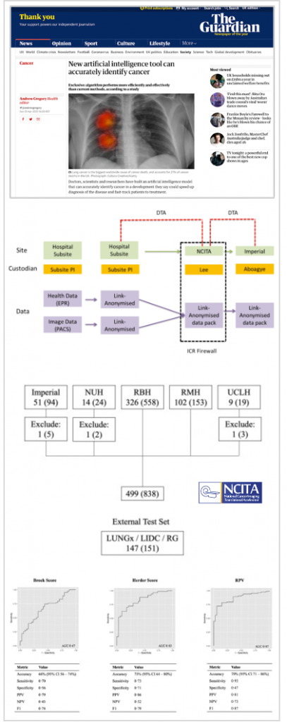
Over the years many different guidelines have been developed to “score” nodules, as seen on imaging, in order to decide how patients should be managed. In the UK, the British Thoracic Society guidelines use two measures, the so-called Brock and Herder scores, but both of these require a large number of clinical variables and the Herder score also needs the patient to have a PET scan. Assembling these variables is time-consuming and so the Lung Nodule Imaging Biobank for Radiomics and AI Research (LIBRA) study (NCT04270799) set out to investigate whether machine learning methods could accurately classify large lung nodules according to cancer risk using only CT data.
XNAT was chosen for use in this study because of its abilities to accumulate images from multiple sites and then make them available to appropriately authorised staff at different institutions. The XNAT Team has longstanding expertise in providing standards-compliant anonymisation of DICOM image data and worked closely with PSs and hospital PACS teams at ten different NHS institutions to source almost 2400 patient imaging sessions (of which only a subset was used in this publication).
Researchers first used the data to create an automatic AI segmentation model to extract lung nodules from the CT images. They then used these regions-of-interest to train a radiomics algorithm to classify the nodules as benign or malignant. In parallel, the Brock and Herder scores were calculated and three radiologists also “read” the scans and predicted the nodule status.
The outcome was assessed by calculating a metric called the Area Under the Curve of the Receiver Operator Characteristic (AUROC). The closer AUROC is to 1, the better is a method’s predictive ability. In this study, the new radiomics model scored similarly (or slightly better) than the human radiologists, and results were also better than the Brock and Herder tests. By modelling a possible decision-support scenario, the research team showed that this method could lead to earlier intervention for malignant nodules that could potentially save lives through earlier interventions in the future.