Research powered by XNAT: RADSARC-R
XNAT is an advanced management system for research images. It uses multiple technologies to provide researchers with the tools they need to process data and view data. The ICR has been at the forefront of developing new capabilities for XNAT, in particular, the ICR-XNAT-OHIF viewer (see Doran et al. Tomography 8.1 (2022). The XNAT Team has the privilege of working with outstanding scientists both locally and internationally, and our tools help them to deliver high-impact clinical studies. This page describes work by the Institute of Cancer Research and Royal Marsden recently published in Lancet Oncology and led by Prof Christina Messiou.
Amani Arthur; Matthew Orton; Robby Emsley; Sharon Vit; Christian Morland-Kelly; Dirk Strauss; Jason Lunn; Simon Doran; Hafida Lmalem; Axelle Nzokirantevye; Saskia Litiere; Sylvie Bonvalot; Rick Haas; Alessandro Gronchi; Dirk Van Gestel; Anne Ducassou; Chandrajit P Raut; Pierre Meeus; Mateusz Spalek; Matthew Hatton; Khin Thway; Cyril Fisher; Robin Jones; Paul Huang; Christina Messiou. Radiomics in sarcoma of the retroperitoneum (RADSARC-R): A novel externally validated CT-based radiomics model for the prediction of histological subtype and tumor grade Lancet Oncology
FEATURED PUBLICATION: RADSARC-R study
RADSARC-R made the headlines on the Today programme. The BBC’s Medical Editor, Fergus Walsh, explains more about the significance of this study.
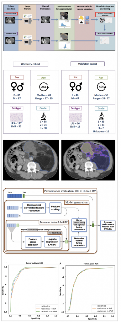
Retroperitoneal sarcomas (RPS) are tumors with unaddressed challenges and a dismal prognosis. Upfront characterisation of the tumor is difficult, and undergrading is common. Radiomics has the potential of non-invasively and globally characterising the “radiological phenotype” of tumours. We aimed to develop and independently validate a radiomics model for the prediction of tumor subtype and grade, based on computed tomography images, for patients with retroperitoneal leiomyosarcoma (LMS) and liposarcoma (LPS).
XNAT was used to assemble a retrospective discovery cohort, containing 170 eligible patients from our tertiary referral centre, and an independent validation cohort of 89 from patients recruited in the EORTC-sponsored STRASS I study. Using the discovery dataset, a radiomics workflow was developed that included manual delineation using the ICR XNAT-OHIF viewer, sub-segmentation, feature extraction and predictive model building, including repeatability testing.
The work resulted in a probabilistic classifier for prediction of sarcoma subtype with an area under the receiver operator characteristic curve (AUROC) of 0.928, and a second predictor of low versus intermediate/high grade (grade 1 versus grade >1) with AUROC 0.882.
To our knowledge, this is the largest RPS cohort that has been analysed by radiomics and the only one that has been validated in an independent cohort. We anticipate that this study will drive further work on integration with other datasets including molecular or digital pathology and the combination with other prognostic tools. This excellent performance could have significant implications in improving accuracy of diagnosis and risk stratification of RPS patients.
For more press reaction to these exciting discoveries, navigate to:
AI and Cancer Diagnosis – “An absolute game changer” – BBC Sounds
AI better than biopsy at assessing some cancers, study finds – Guardian
Scientists excited by AI tool that grades severity of rare cancer – BBC News
Prof Christina Messiou interviewed by Amol Rajan – BBC Radio 4 Today